Wojcik, Gabriela MSc; Parekh, Mohit PhD; Romano, Vito MD; Ruzza, Alessandro MSc; Scorcia, Vincenzo MD; Viola, Pietro MD; Leon, Pia MD; Franch, Antonella MD; Gadhvi, Kunal A. MB BS; Ponzin, Diego MD; Ferrari, Stefano PhD
Author Information:
International Centre for Ocular Physiopathology, Fondazione Banca degli Occhi del Veneto Onlus, Venice, Italy;
Department of Ophthalmology, Schepens Eye Research Institute of Massachusetts Eye and Ear, Harvard Medical School, Boston, MA;
Clinical Eye Research Centre, St Paul’s Eye Unit, Royal Liverpool University Hospital, University of Liverpool, Liverpool, United Kingdom;
Department of Medical and Surgical Specialties, Radiological Sciences, and Public Health, Ophthalmology Clinic, University of Brescia, Italy;
Department of Medical and Surgical Sciences, Cornea and Ocular Surface Unit, Magna Graecia University of Catanzaro, Catanzaro, Italy;
The Ophthalmology Operational Unit, Structural Department Specialist Surgery Vicenza, San Bortolo Hospital Vicenza, Vicenza, Italy; and
Department of Ophthalmology, Hospital SS. Giovanni e Paolo, Venice, Italy.
Correspondence: Mohit Parekh, MSc, PhD, AFHEA, Department of Ophthalmology, Schepens Eye Research Institute of Mass Eye and Ear, Harvard Medical School, Boston, MA 02114 (email: mnparekh@meei.harvard.edu).
The study was partially funded through the 5X1000 funds to Fondazione Banca degli Occhi del Veneto from the Italian Ministry of Health and the Italian Ministry of University and Research.
The authors declare no conflict of interest.
G. Wojcik and M. Parekh have contributed equally.
Cornea 43(1):p 38-44, January 2024. | DOI: 10.1097/ICO.0000000000003274
Abstract
Purpose:
The objective of this study is to validate Descemet membrane endothelial keratoplasty (DMEK) Rapid device for preloading DMEK grafts with endothelium outward.
Methods:
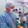
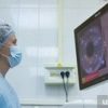
In this multicenter retrospective clinical study, DMEK tissues (n = 27) were peeled and preloaded (8.25 mm) in a DMEK Rapid device. The device was loaded in a container prefilled with the storage solution and shipped from a single center in Italy to 4 different centers located in Italy and the United Kingdom. Preloaded tissues were delivered by injecting the graft in the anterior chamber. Patients were monitored at days 1 and 15 and at months 1, 3, and 6, as well as at the last follow-up (9–12 months) postoperatively. Main outcome measures included rebubbling rate and graft failure, corrected distance visual acuity, endothelial cell loss (ECL), and central corneal thickness at all time points. A one-way analysis of variance test comparing day 1 with all later time points was followed with significance at P < 0.05.
Results:
The average recorded surgical time was 6 to 25 minutes with no immediate surgical complications. Rebubbling was observed in 7 of 26 cases with one graft failure within 15 days postoperatively. The mean corrected distance visual acuity at day 1 was 0.64 ± 0.49 logMAR, which improved to 0.18 ± 0.43 logMAR at the last follow-up. Endothelial cell density values showed a significant decrease at the last follow-up (1827 ± 565 cells/mm2) (P < 0.001) compared with the preoperative value (2503 ± 128 cells/mm2), with an average endothelial cell loss of 27%. Central corneal thickness significantly dropped from 694 ± 157 μm at day 1 to 502 ± 42 μm at the last follow-up (P < 0.001).
Conclusions:
DMEK Rapid device is quick, easy, and efficient for preloading and shipping DMEK grafts internationally in endothelium-outward orientation.
Copyright © 2023 Wolters Kluwer Health, Inc. All rights reserved.